The skeleton known as Little Foot was first discovered in 1994 in the limestone caves of Sterkfontein in South Africa. However, the skull of this fossil suffered significant damage and deformation due to geological pressure, making it difficult to study using traditional methods. In 2019, researchers transported the skull to the Diamond Light Source synchrotron facility in the UK for high-tech scanning. After that, scientists used semi-automated methods and supercomputers to separate bone fragments, resulting in a 3D reconstruction with a resolution of 21 microns.
“We have made significant progress in creating a quality reconstruction, something that was not achievable with the physical remains,” noted the lead author of the study, Amélie Bode, a paleoanthropologist from the PALEVOPRIM laboratory associated with CNRS in France.
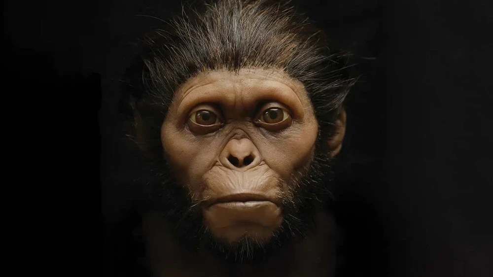
The reconstructed face features broad eye sockets and a morphology reminiscent of East African australopithecine samples, in contrast to findings in South Africa. This discovery raises questions about the migration of early hominids; one hypothesis, as reported in Science News, suggests that Little Foot may have belonged to a group that migrated from East Africa to South Africa over 3.5 million years ago.
However, Bode cautioned about the need for caution, as the number of available australopithecine skulls for comparison is limited. “We have only a few samples, so any conclusions must be made with caution,” she added in an interview with Science News.
The team plans to expand the digital reconstruction to include other parts of Little Foot's skull, such as the braincase and teeth. This may aid further research concerning the diet, biomechanics, and brain evolution of early hominins. The 3D reconstruction will be available to the scientific community, allowing for additional studies.


